Microanalysis has recently acquired an Agilent 7890 GC-MS (Gas Chromatogram – Mass Spectrometer) for its new premises at East Perth.
The GC-MS will expand Microanalysis Australia’s capabilities in the areas of material identification and characterisation further into the organic sphere, specifically in the areas environmental analysis, polymer characterisation geochemical and industrial chemistry analysis.
This will complement our existing strengths in the areas of mineral and inorganic identification using scanning electron microscopy, light microscopy and x-ray diffractometry.
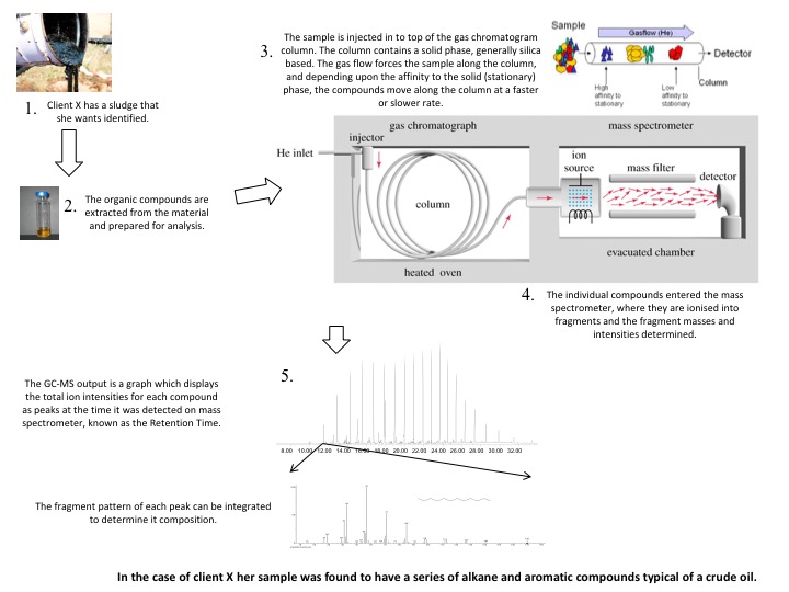
A GC-MS is composed of three basics building blocks:
A means of introducing the sample via an injection port.
The separation of mixtures of compounds by partitioning them between a gas phase (typically helium) and an activated solid phase attached to a silica capillary column. The difference in the chemical properties between different molecules in a mixture and their relative affinity for the stationary phase of the column will promote separation of the molecules as the sample travels the length of the column. The molecules are retained by the column and then elute (come off) from the column at different times (called the retention time).
As the helium gas phase containing the molecules passes through the mass spectrometer, they are ionised into fragments and detected based on their mass charge ratio. The fragment pattern can be matched to an internal library to provide the absolute identification of a compound.
The true power of the GC-MS is when the information of the retention time and mass fragment patterns are combined to allow the identification of compounds in extremely complex samples such as crude oils, as well as enabling the detection of pesticides, PCB and PAHs down to ppb or even ppt levels.
The diagram below provides a summary of how a typical sample would be analysed on this system.
MAA is planning to develop its investigative and analytical base over the coming months to provide clients with specialised services in the areas of organic analysis.
Be sure with Asbestos
We often think of asbestos as being something from a bygone era which isn’t compatible with today’s health standards. Sadly, the prevalence of asbestos containing materials (ACMs) worldwide is still stubbornly high¹. As well as the ‘70s buildings needing repair or being demolished, there has been an increase in incidents of new imported products being found to contain asbestos. Importing asbestos at any concentration is illegal in Australia².
Asbestos can be found in a range of building materials such as cement, plasterboard, vinyl, window putty and roof tiles, as well as consumer products like crayons, paint and baby powder.
When to suspect asbestos
Asbestos containing material isn’t always obvious. It is worth checking whether a product contains asbestos if any of the following are true:
The product was manufactured in the 1960s, 1970s or 1980s;
There are visible, very fine white, blue, green or brown fibres protruding from the product surface or from broken edges of the product;
The product is plasterboard, fibreboard, vinyl, rubber, wax, cement sheeting or other building material and was recently imported; or
The product has any evidence of fibrous, non-flammable dust.
What to do if you suspect you have asbestos
If you think you may have an ACM, make sure you keep it moist to prevent the asbestos fibres from becoming airborne. This will prevent the asbestos from entering your lungs.
Put a small section (~ 5 cm x 5 cm) in a plastic, sealable bag such as a zip lock bag. Place this bag inside another plastic bag.
Now you’re ready to take your sample to a lab for analysis!
What testing is available
Currently, polarised light microscopy, or PLM, is the first line analysis for detection of asbestos. Over the years there have been several areas of concern raised about the detection limits and technical limitations of the technique. PLM involves examination of the sample using an optical microscope to identify whether fibrous material is present and it is able to differentiate several different types of asbestos.
Scanning Electron Microscopy, or SEM, is an alternate technique to identify asbestos using newer technology. SEM provides quick, accurate detection in a wide variety of different materials. Using Energy Dispersive X-ray Spectroscopy (EDS) with SEM imaging, the elemental composition can be determined to easily and conclusively differentiate between different types of asbestos and non-asbestos fibres (eg fibreglass). SEM EDS also has a much lower detection limit than PLM, often to better than 0.001 wt %, of detecting fibres as small as 50nm in diameter and detecting in a wider background of materials such as in or on ores, aggregates, fillers and polymers.
A recent bulletin³ by the Department of Mines and Petroleum (DMP) outlines the importance of detecting these very fine fibres as they are more likely to move into the deeper parts of the human respiratory system and lodge in the lungs, causing cell damage. The DMP bulletin explains that asbestos fibres below the resolution limit of PLM are easily detected and identified by SEM EDS.
We have outgrown our much loved space in Victoria Park and have acquired a new property in East Perth. The move is taking place Friday, 22 July 2016.
Our new laboratory is located at: 37 Kensington Street, East Perth
Decommissioning of our instruments will take place on Thursday, 21 July 2016 and recommissioned over the weekend. If you have an urgent job on its way to us please contact Rick Hughes on 0407 771 447.
We will be operating from East Perth as of Monday 25 July 2016, please amend your records to account for our change of address.
Our phone number will remain +61 8 9472 4880 and after hours 0407 771 447
SEM Image Auction
Start your bidding…
Microanalysis have pledged to support the Starlight Children’s Foundation who have been helping children and their families since 1988.
These beautifully unique SEM images are being auctioned off by the team at Microanalysis. Professionally printed (12 inch x 12 inch) and framed they would make a stunning addition to your office or home, and will raise some well needed funds to support the incredible work of the Starlight Children’s Foundation.
Forensic Analysis of Errant and Nuisance Particles
Many of us have been in the situation where we look at our car or our washing and wonder ‘just how did it get covered in that dirt?’ In most cases nuisance particles are just that, ‘a nuisance’, causing time and effort/cost to remove or clean. In some cases these same particles can be quite harmful, in others it can mean a breach of compliance with local, state or federal government regulations which limit or control the emissions that are allowed to be released from a business or process.
So how can you tell what a particle is and where it’s come from?
Microanalysis Australia is routinely tasked to do just that. Metal fabrication plants often grind, weld, sand blast and spray paint as part of their daily manufacturing/repair processes. It may not always be possible to conduct such activities in a controlled (indoor) environment and occasionally a release of errant particulate may be carried on the wind onto neighbouring properties. Depending on the wind conditions, freshly created particles of metal fragments, welding fume or spray paint may drift and settle on buildings, cars, boats etc which, if left unchecked can build up to unsightly levels at best or damage surfaces prematurely and cause ill health at worst.
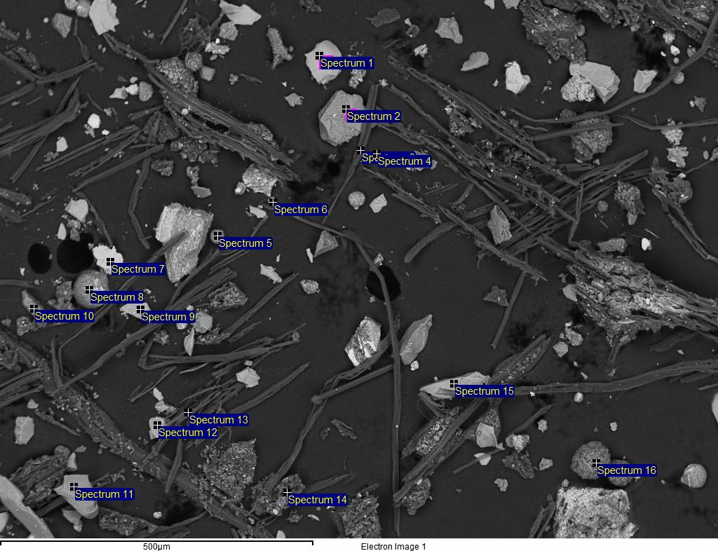
Scanning electron microscopy (SEM) allows the automated or manual, rapid analysis of between hundreds and thousands of particles and the associated mineralogy and likely origin to be determined.
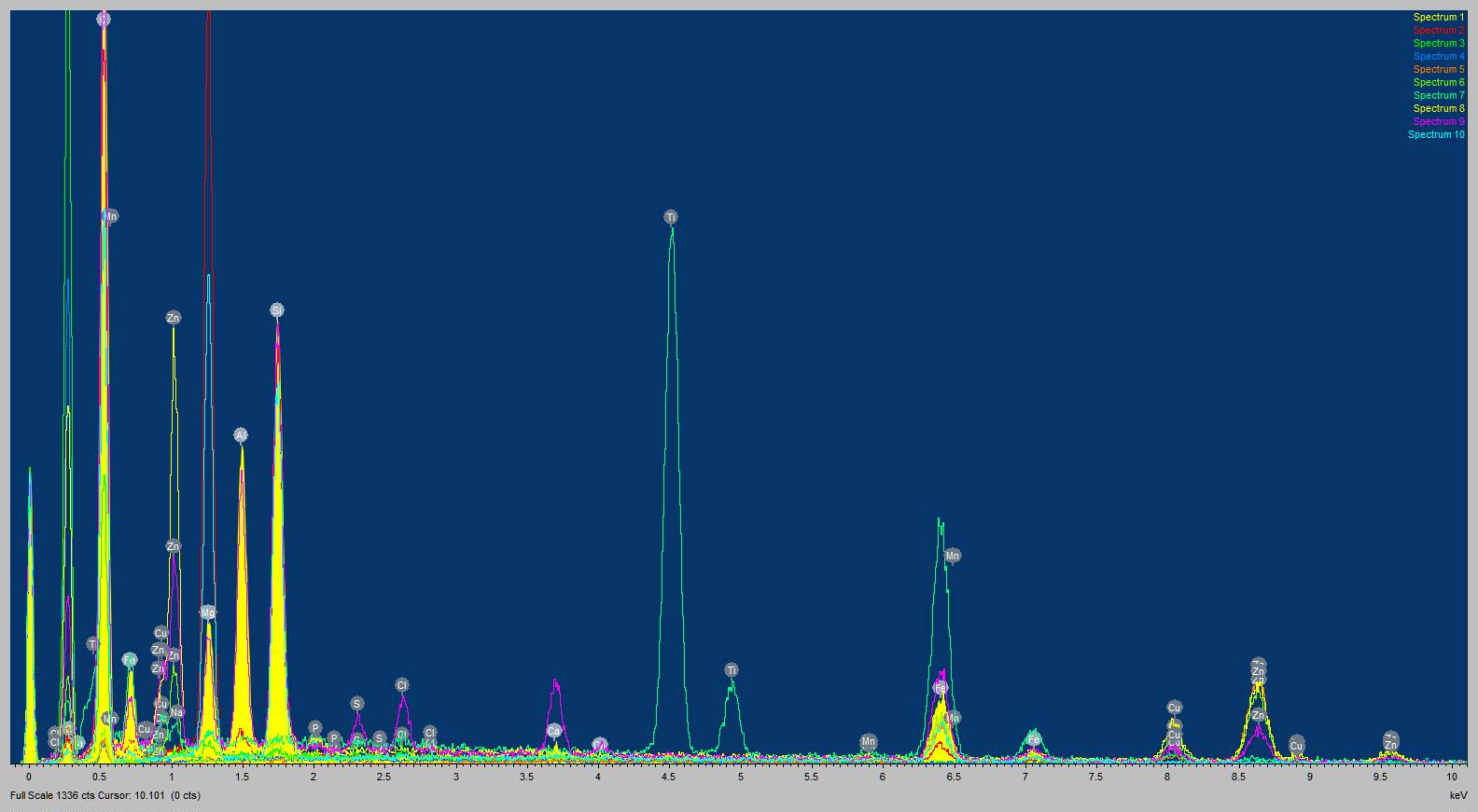
The particles in the above image were collected on site using double-sided carbon tape, an SEM-ready acquisition technique that allows particles to be quickly analysed and interpreted. The particles range in size from 5 µm to 80 µm and represent garnet blast media covered in paint and steel fragments from a metal preparation process. Carbonaceous fibres from local flora can also be seen. The relative abundance of the elements can be used to compare against suspect mineral/metal phases and used to positively identify particles by specific origin.
Microanalysis can differentiate sand blasting media – from garnet, olivine, staurolite and other neosilcate minerals, to metal/metal oxides, glass beads and steel media. Tell-tale signs of controlled particle size coupled with high angularity (post blast-induced fracture) and associated pigments such as titania and barite lend weight to the activities being conducted when the blast media was being used. Welding fume has its own size, morphology and chemical fingerprint and is readily distinguished from background environment dust.
Local regolith (local environment geology) often needs to be taken into consideration when highlighting anthropogenic phases.
Airborne dusts from stockpiles or earthworks may travel considerable distance if their size distribution is fine enough. Naturally occurring mineral fibres or components of low toxicity at the point of origin may be concentrated and effectively refined by wind transport meaning that locations downwind may be exposed to a size range and composition that may only represent a trace component of the original material. Air monitoring is useful in abstracting inhalable and respirable size fractions over longer acquisition times to get time weighted averages of dust emissions for both static (positions in a designated environment) and personal (individuals moving around an environment). Microanalysis routinely measures the quantity and composition of particles on filters used to collect both total suspended particulate (TSP) and inhalable (PM10) and respirable (PM4) particles. Morphologies and mineralogies of these particulates are crucial in understanding the risks posed to health as well as general amenity.